


From childhood we get used to running, jumping, guys like to climb and play football, girls in ropes and more.And the active lifestyle is thus entering the human mind that over the years, when the muscles pulled out somewhere, somewhere the joints is ill, the person doesn't even pay attention - "Well, think how many times the boiler hurts."Here in today's article, talk and why the knee can injure, and is it always the usual outcome of sharp movements.
What is arthrosis?
Arthrosis -In the group of diseases of the muscle-bone system of various origin, but with similar biological, morphological and clinical events.The basis of their development is a degenerative lesion of all components of the joint, primarily cartilage, thin bone, sinovial membrane, ligaments, capsules and periarticular muscles, with the formation of marginal osteophytes and pure or hidden moderately expressed sinovitis.Because with this disease of pathological change, they capture both cartilage and bone tissue.
Arthrosis is often referred to osteoartostAnd sometimes osteoarthritis.
Statistics (epidemiology)
Among all the diseases of the arthrosis joints is up to 80% of cases.
The disease develops mainly in central and age.In a young age, the arthrosis can appear after injuries of joints, inflammatory processes, as well as in the congenital pathology of the musculoskeletal system.
X -Ray signs of arthrosis are revealed in most people older than 65 and almost 95% older than 70 years.
Women suffer from arthrosis almost twice more often than men.The incidence rate increases during postmenopause.
The great role in the development of arthrosis play hereditary factors.It was found that the frequency of disease development in the families of patients with osteoarthritis is twice as much as possible, and the development of arthrosis in innate shortcomings of the muscle-bone system increases by 7-8 times.
Arthrosis - ICD
- MKB-10: M15-M19, M47
- MKB-9: 715
- MKB-9-km: 715.3
Symptoms of arthrosis (clinical picture)
Clinical manifestations of the disease and their seriousness depend on the localization of the pathological procedure, the health condition of the patient and the image of his life.
The first signs of arthrosis
Arthrosis often begins gradually, imperceptibly for the patient.
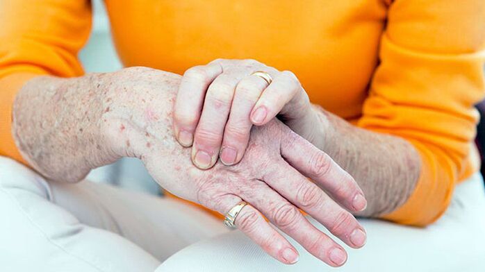
The first symptom of the disease is usually short-minor joint pain (arthralgia), which has the greatest load.These are, above all, the joints of the lower extremities - knee, hip, plus-falanx compounds of the first thumb foot.From the head of the upper limb, interfalgeal compounds often affect the brush blend for carpal pattern brushes.
Arthrosis usually starts with a lying one wrist, but after a while and other compounds are included in the process.
The main symptoms of arthrosis
With arthrosis, patients complain about pain, crumbly, movement limit in common, swelling and joint deformation.
Separately, it is worth the abiding nature of pain.It is possible with arthrosis, mechanical and initial pain.Mechanical pain occurs with a load on the affected joint.Such pain is tormented mostly in the evening at rest, disappears after a few hours of rest.The appearance of this type of pain is associated with gradual increase in bone pressure during physical effort.The pressure causes bone beams and irritation of boning tissue.
The initial pain appears at the beginning of a walk, then stops quickly and appear again during physical effort.Starting pain can occur with the friction of the joint surfaces of the affected joint.Small particles of necrotic cartils fall on the surface of the cartilage.At the first steps and the particles are pushed into the cavity of the common bag and the pain ceases.
In addition to arthrosis, pain can be associated with periarthritis and teodo (inflammation of soft periarticular tissues, ligamental apparatus and articulated bags).This pain only occurs during the movements in which affected tendons participate, as well as in certain joint positions during the movement.
Pathological changes generally begin large compounds that are exposed to great physical effort during the day.At the beginning of the disease, the pain occurs as a result of the inconsistency of the possibility of microcyirculatory channel with the needs of articulated tissues.Therefore, they will reduce pain, patients slowly take the first few steps and only then speed up the pace of walking.Pain can appear after half to two hours to walk or work in a standing position.This is a signal to change load, backrest or job type.
In the later stages of the disease, arthralgia can appear with minimal joint loads and remain at rest for a long time.This is due to the fact that in later stages, rude changes in common tissues, destroying articulated cartilage and secondary synovitis are formed.Development of massive, gross change in bone bone tissue, its individual fragments can be separated and, fall into a common gap, cause harsh pain.This phenomenon is called a symptom of an articulated mouse.
During the examination of the joints, the deformation was observed.In addition, with arthrosis there is a thickening of ferry soft tissues, the hippotrophy of regional muscles, movements of the axis of the limbs.The thickening of the interfalangeal joints with bone growth and periarticular fabrics seal is called Gerberden nodes.
Pain when they feel sharply localized in a joint jaith, a shared capsule attachment places, but this symptom of the disease is not always.Swelling and joint pain is determined by secondary sinovitis.
Breach of a common function in early stages of arthrosis is manifested by the restriction of the amplitude of the movement.This is due to the lesion of perhetic tissues and sinovitis.
In the later stages of the disease, the clinical manifestation of the contract develop different in terms of seriousness.The most common is the functions of the knee and hip joints.
Symptoms of arthrosis Depending on the localization of pathology
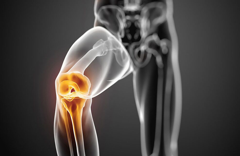
Arthrosis with knee joint damage - Symptoms
The lesion of the knee joint with arthrosis is called gonartrosis.Primary gonortiness is developing in menopausal women.The reasons for the mean are the most common injuries of a common knee and violating statics with curved vertebrae, straight legs.Patients complain about the pain in the knee joint that occur during the movement, especially when you walk the stairs.The pain is localized on the front or interior of the knee wrist.The jig movements are limited: first bending and later extension.The occasion is moving, the crisis often appears.The development of reactive sinovitis, pain during the movement is intensified and cared at rest.Ankling swelling, pain during palpation, redness (hyperemia) and increasing the skin temperature are determined.Over time, due to bone growth, the knee joint deformations occurs.
Arthrosis with hip joint damage - Symptoms
The hook joint lesion is called coxartrosis.This is the hardest form of arthrosis.The causes of the disease can be inborn dysplasia hip joints, injuries, menopause.Patients have joint pain during the movement, in a standing position.The constraint of the joint movement is gradually increasing (first internal and external rotation, later flexion).There is a christ associated with limb shortening.With bilateral damage, the duck walk is typical.Atrophy of thigh muscles and buttocks is developed.There is no joint swelling with Catros.Palpation determines limited pain in the female head.
In the initial phase of arthrosis, joint functions are preserved.With further disease development, it is first temporarily limited, and then the ability to work is completely lost, the patient loses the ability of the supermarket, external assistance.
Causes of arthrosis
Arthrosis is based on the primary degeneration of the articulated cartilage with accompanying destructive changes in the bones that make up a wrist.Such degeneration arises as a result of imbalance between mechanical loads on the cartilage coupling and the possibility of fees for this load.
In the development of degenerative changes in articulated cartilage, several factors can participate in the same time:
- Functional overloads, including professional, households and sports, causing cartilage mycotrauum;
- Joint injuries;
- Contagious and unnecessary joint inflammation;
- Joint dysplasia, leading to a violation of the comparison of common areas;
- Violation of body statica as a result of the curvature of the spine (Kifoza, Scoliosis, Pathological Locose, etc.), flat feet;
- Chronic hemartrosis:
- Diseases with metabolic disorders (gout, obesity, chondrocalcinosis);
- Osteodistrophy or peget disease;
- osteomyelitis;
- Pathology of peripheral nervous system with loss of sensitivity;
- Endocrine pathology (acromegaly, diabetes, amenorrhea, hyperthyroidism);
- Hereditary tendency.
Arthrosis risk factors include older people, female sex, obesity.
Development mechanism
Metabolic disorders in cartilage are based on quantitative and qualitative changes in the main substance of the cartilage.The main substance consists of prostelogues that provide collagen stability.The development of the arthrosis is followed by insufficient formation or increased destruction of cartilage components.
With osteoarthritis in the cartilage tissue, hyaluronic acid content, chondroitine and keratin decreases.In addition, modified proteoglycans lose the ability to retain water.A collageni is absorbed that makes it difficult, causing the decrease in resistance to cartilage.
If chondrocytes are damaged, they begin to produce collagen and proteoglycane that are not characteristic of normal cartilage tissue.These modified substances cause the loss of biochemical quality cartilage.
Of great importance in the development of arthrosis are immune disorders.Destruction of proteobican cartilage monitors the appearance of immune reactions of mobile and humoral type.In return, this causes progressive fibrosis and sclerosis of the sinum membrane, pathological changes in intraunticular synovial fluid and cartilage violation.The inferior Sinovian shell supports the progress of degenerative changes in the articulated cartilage.
The hereditary factor has a certain value in the development of arthrosis.
Classification of arthrosis
Arthrosis is divided into two groups: primarily and secondary.
In distribution (primary arthrosis):
- Locally (with a three-joint impairment)
- common or generalized, polyarthrosis (defeat three joints or more).
Depending on the destination (secondary):
- A. Tasobed wrist (KokeSartrosis);
- A. Common knees (gonartrosis);
- A. The wrist of the elbow;
- A. The blend of shoulder;
- A. skins;
- A. Cervical Department (Unkoartrosis);
- A. Hands;
- A. Ankle wrist (cruzartrosis)
- A. Stop.
According to the etiology:
- Post -traumatic
- metabolic
- Due to endocrine pathology.
Diagnosis of arthrosis
The diversity of clinical events and arthrosis variants makes it difficult for the early diagnosis of the disease.The falsity of the diagnosis is also associated with the lack of certain symptoms, hidden starting of the disease.Of great importance, the definition of factors contributing to the development of arthrosis:
- chronic articulated trauma;
- Long time execution of stereotypical movements;
- Physical activity on the compound for a specified time;
- a violation of salt or fat metabolism;
- Hereditary vices of a muscle-bone system.
The X -Ray examination is the most important meaning in diagnostics of arthritis.Radiography of viewing both knee joints is performed in a direct position, bent position, next to the lateral position.Classic signs of arthrosis on the radiograph are: narrowing a joint gap, the presence of osteophytes, subhondral sclerosis and subhongral cysts.There are following phases of radiological changes in arthritis:
- 0 - No change.
- And - Radiological suspicious signs.
- II - Minimal changes (slight narrowing of the shared gap, Osteosclerosis branch, individual osteophytes).
- III - moderate events (moderately narrowing of the Charter, more osteophytes).
- IV- Expressed changes (joint gap is not visible, the multiple rude osteophytes is determined), the synonitis is often present.
In the presence of these symptoms, further tools are not needed.
In their absence or low weight, the joints, MRI, scintigraphies were performed.
Clinical blood, urine and intraarticular fluids are not included in the list of mandatory studies for diagnosis of arthrosis.But these tests are necessary to exclude such articular pathology.
Main clinical and diagnostic signs of arthrosis:
- Mechanical pain in the joint;
- fatigue;
- a sense of instability in the joints of the lower extremities;
- Damage to the joints of the first finger and arm;
- gradual start of the disease;
- slow progressive current;
- Common deformation;
- Hypotrophy of regional muscles;
- Repeating sinovitis;
- Limitation of the motion in the joint;
- X -Ray changes.
Arthrosis must be distinguished with damage to the compounds with rheumatoid arthritis, contagious, metabolic and reactive arthritis.
Rheumatoid arthritis, unlike arthrosis, starts inflammation of small joints of hand and foot.It is characterized by intense pain in the inflammatory type, morning joint rigidity, the presence of rheumatoid nodules.
Gotric Arthritis is located mainly in men.A high local activity with acute paroxysmal pain in the first plus-phalan body blend is characteristic.With Gout, the presence of Tofus is typical, there are "punch" on the radiography.
Psoriatic arthritis characterize skin lesions, especially scalp, finger deformation in the shape of spindles and bright color of raspberry skin above affected joints.
Earned arthritis is characterized by acute start, rapid development and course, sharp pain, high temperature and efficiency of antibacterial therapy.
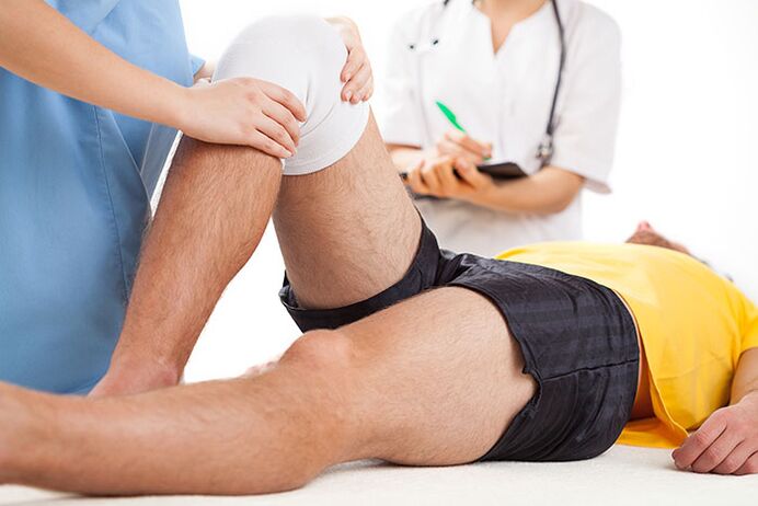
Treatment of arthrosis
The treatment for arthritis should be long, complex.Basic principles of arthrosis treatment:
- Unloading of compounds (correct manner of mobility and mechanical loads, dosed walking, reducing body weight, long-term standing, weight wear, strengthening muscle-ligament devices, massages, electrical stimulation).
- Conservative correction of static disorders (use of orthopedic shoes, corsets, supervisors).
- Impact on total metabolism and blood circulation (use of biostimulants, vasodilation remedies, balneotherapy and physiotherapy courses twice a year).
- Elimination of reactive sinovitis, antinamental therapy.
Arthrosis patients show nutrition with salt, sugar, strong tea, coffee, smoked meat, sharp dishes.This improves the sensitivity of vascular and articulated receptors, returns the blood vessel tone, normalizes the exchange in chondrocytes.With arthrosis, it is necessary to drink enough liquid (at least 8 glasses of water a day).
Drug treatment The arthrosis includes the use of rapid -acting anti -infalmaturine and side remedies (non-steroidal anti -infalmalmic drugs - NSAIDs), basic medications - hondroprotectors.Non-?Nonceptive and selective TSO-2 inhibitors are used from the NSAID.
As local therapy for affected joints are used by NSAIDs in the form of fat or gel.
In the presence of reactive sinovitis, tendinitis or tendovaginitis, when the NSAID treatment is inefficient, appropriate intraArticular or intramuscular application of corticosteroids.
Basic therapy with hondroprotectors (hondroitin, glucosamine, hyaluronic acid) Used to prevent the articular brisker degeneration.
Hondroprotektor treatment is indicated in the clinical and radiological phases of the I-III arthrosis.
In addition to direct hondroprotects, drugs that encourage the reconstruction of cartilage (biogenic stimulants).These drugs are used during the remission, in the absence of reactive sinovitis.
Arthrosis, medications that improve microcirculation are also marked.In the presence of varicose veins of the lower extremities, a correction of venous blood flow is needed.
In patients with arthrosis, it is necessary to diagnose in a timely manner and treat osteoporosis.
Arthrosis physiotherapy
Physical methods of treatment also refer to the basic arthrosis therapy.According to their influence, metabolic processes, microcirculation of blood and tissue fluid, is renovated neurogumal regulation.
The treatment complex arthrosis includes induction, microwave therapy, pulserene electricity, drug electrophoresis and magnetotherapy.To remove the sinovitis, ultraviolet radiation areas of affected joints in erythemic doses, the electric field of Ultra -high frequency, electrophoresis with anal, dimexide or hydrocortisone.
To prevent arthrosis advancement, it is recommended to reduce body weight, avoid increasing loads on the joints, walking in suppression, increased humidity and hypothermia.An individual choice of shoes and supervisors is important.
It has been shown to strengthen regular physical exercise, swimming, cycling to strengthen muscles.Classes of heavy and light athletics, football is not recommended.
Therapeutic exercises is performed differently, in a seated position, lying, in the pool.The movements should not be intensive, traumatic, their scope and number of repetitions increase gradually, avoiding overload.
Popular and efficient methods of treatment arthrosis include massage and kinesitherapy.
With significant changes in deformation joints, mobility limit, surgical treatment is recommended.Arthroplasty, endoprosthetics, osteotomy are performed.
Forecast of diseases
Primary arthrosis rarely leads to complete disability.In the presence of reactive sinovitis, patients temporarily became disabled, and sometimes they are forced to change the profession.With secondary crachija, the forecast is less favorable due to the rapid progressive flow of diseases with the development of significant impaired joint functions.In such cases, the disability may occur over several years of the disease.
Arthrosis prevention
The primary prevention of arthrosis should be started in childhood.That's the following:
- Prevention and treatment of scoliosis;
- Straightening straight feet using special supervisors;
- Physical education classes for strengthening muscles and ligaments;
- Rational nutrition and prevention of metabolic disorders;
- Limitation of severe sports in childhood and adolescence;
- alternating work sitting at a walking table;
- Correct organization of work and the rest of employees in companies where there is great physical activity.
Secondary prevention provides for measures that prevents the development of repetitive reactive Sinis.These include dosed walking, limiting physical effort, support with support and other measures that unload joints.With heavy symptoms of arthrosis, it is necessary to constantly download basic medications.General strengthening therapy, improving blood circulation and metabolism, the annual treatment of the spa is recommended.
Which doctor will he go to?
- Rheumatologist
- Orthopedist